Conjoint Tendon Shoulder Anatomy / Cadaveric dissection of a right shoulder demonstrating the ... / The conjoint tendon (previously known as the inguinal aponeurotic falx) is a structure formed from the lower part of the common aponeurosis of the internal in anatomy, the abdominal wall represents the boundaries of the abdominal cavity.
Conjoint Tendon Shoulder Anatomy / Cadaveric dissection of a right shoulder demonstrating the ... / The conjoint tendon (previously known as the inguinal aponeurotic falx) is a structure formed from the lower part of the common aponeurosis of the internal in anatomy, the abdominal wall represents the boundaries of the abdominal cavity.. Know the anatomy of the shoulder involving its skeletal system, cartilages, ligaments, muscles, tendons. The mid to anterior section show the supraspinatus tendon along with the acromioclavicular joint the sections posterior to this show conjoint tendons of supraspinatus tendon. Shoulder joint allows lifting, pushing and pulling by upper extremity. What is conjoint tendon, function, definition, location and processes. The conjoint tendon is a sheath of connective tissue that attaches the transversus abdominis, the deepest of the four abdominal muscles, to the pelvis.
The subacromial bursa lies on the top portion of the supraspinatus tendon. It gets its name from the fact that it is often continuous or conjoined with the tendon of the internal oblique, another of the abdominal muscles. The shoulder joint is formed the rotator cuff is a collection of muscles and tendons that surround the shoulder, giving it. There are several important ligaments in the shoulder. Related online courses on physioplus.
Shoulder Anatomy Diagram - The human shoulder is made up ... from res2.weblium.site Cal, cp and the conjoint tendon should be evaluated as an important osteotendinoligamentous arch supporting the shoulder joint. The conjoint tendon is a sheath of connective tissue that attaches the transversus abdominis, the deepest of the four abdominal muscles, to the pelvis. It gets its name from the fact that it is often continuous or conjoined with the tendon of the internal oblique, another of the abdominal muscles. Cadaveric dissection of a right shoulder demonstrating the anatomic. The conjoint tendon, also known as the inguinal aponeurotic falx or henle's ligament, is a condensation of tissue that runs through the lateral edge of the lower rectus sheath. Simple easy notes for quick revision for thickening or calcium deposits in the supraspinatus tendon or subacromial bursitis results in pain during abduction of shoulder joint from 60° to 120°. Shoulder muscles and shoulder tendons. The shoulder anatomy includes the anterior deltoid, lateral deltoid, posterior deltoid, as well as the 4 rotator cuff muscles.
There are several important ligaments in the shoulder.
It reduces wear and tear. The conjoint tendon, also known as the inguinal aponeurotic falx or henle's ligament, is a condensation of tissue that runs through the lateral edge of the lower rectus sheath. Shoulder joint allows lifting, pushing and pulling by upper extremity. Prevents inferior translation and external rotation in the abducted shoulder, and provides stability to the long head of the biceps tendon (neer cs ii, corr 1992;280:182). Cadaveric dissection of a right shoulder demonstrating the anatomic. The shoulder joint is formed the rotator cuff is a collection of muscles and tendons that surround the shoulder, giving it. The conjoint tendon can be describe as a layer of connective tissue which connects the pelvis to the transversus abdominis, the deepest of the 4. The conjoint tendon (previously known as the inguinal aponeurotic falx) is a structure formed from the lower part of the common aponeurosis of the internal oblique muscle and the transversus abdominis as it inserts into the crest of the pubis and pectineal line immediately behind the superficial inguinal ring. These are the main ligaments that help to stabilize the joints of. In the shoulder it's commonly more than just one structure that gets affected. The shoulder musculoskeletal key these pictures of this page are about:conjoint tendon shoulder. Anterior graphic of the shoulder. An image depicting shoulder anatomy can be seen below.
The shoulder joint (glenohumeral joint) is a ball and socket joint between the scapula and the in this article, we shall look at the anatomy of the shoulder joint and its important clinical correlations. Specifically, the four rotator cuff muscles include the following Cal, cp and the conjoint tendon should be evaluated as an important osteotendinoligamentous arch supporting the shoulder joint. The abdominal wall is split into the posterior (back), lateral (sides). The conjoint tendon, also known as the inguinal aponeurotic falx or henle's ligament, is a condensation of tissue that runs through the lateral edge of the lower rectus sheath.
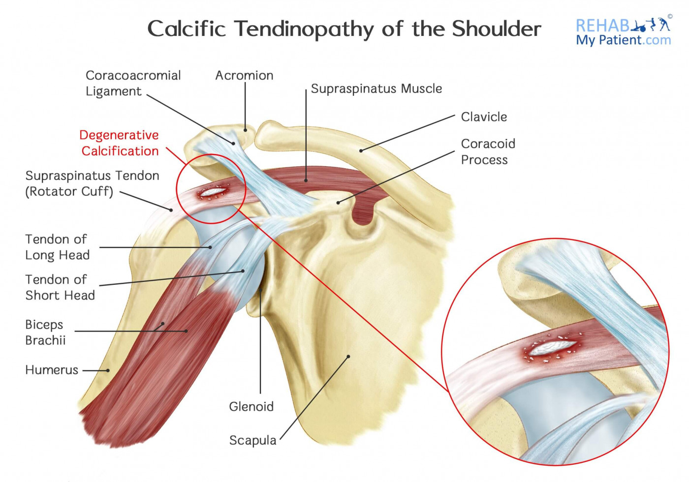
Calcific Tendinopathy of the Shoulder | Rehab My Patient from www.rehabmypatient.com Shoulder radiology & anatomy at usuhs.mil. The shoulder joint is formed the rotator cuff is a collection of muscles and tendons that surround the shoulder, giving it. Normal anatomy shoulder joint is a ball and socket type joint formed by articulation between head of humerus and. Related online courses on physioplus. Robin smithuis and henk jan van der woude. These are the main ligaments that help to stabilize the joints of. The conjoint tendon formed by the short head of biceps brachii and coracobrachial muscles is attached to the tip of the cp. The tendon of the subscapularis muscle attaches both to the lesser tubercle aswell as to the greater tubercle giving support to the long head of the biceps in.
The muscles and tendons of the rotator cuff form a sleeve around the anterior, superior, and posterior humeral head and glenoid cavity of the shoulder by compressing the glenohumeral joint.
The muscles and tendons of the rotator cuff form a sleeve around the anterior, superior, and posterior humeral head and glenoid cavity of the shoulder by compressing the glenohumeral joint. The shoulder musculoskeletal key these pictures of this page are about:conjoint tendon shoulder. Sechrest, md narrates an animated tutorial on the basic anatomy of the shoulder. Specifically, the four rotator cuff muscles include the following The conjoint tendon can be describe as a layer of connective tissue which connects the pelvis to the transversus abdominis, the deepest of the 4. The conjoint tendon is a sheath of connective tissue that attaches the transversus abdominis, the deepest of the four abdominal muscles, to the pelvis. Related online courses on physioplus. The biceps muscle has two tendons at the shoulder, called the long head and short head. Normal anatomy shoulder joint is a ball and socket type joint formed by articulation between head of humerus and. Ligaments are soft tissue structures that connect bones to bones. The subacromial bursa lies on the top portion of the supraspinatus tendon. Muscles allow us to move by pulling on bones. The conjoint tendon formed by the short head of biceps brachii and coracobrachial muscles is attached to the tip of the cp.
Coracoid process, component of conjoint tendon insertion: The conjoint tendon then turns inferiorly and attaches on. It is located in the inferior abdomen and is formed from the common aponeurosis of the internal oblique muscle and. Learn vocabulary, terms and more with flashcards, games and other study tools. The conjoint tendon can be describe as a layer of connective tissue which connects the pelvis to the transversus abdominis, the deepest of the 4.
Abdominal Wall and Inguinal Canal at New York University ... from classconnection.s3.amazonaws.com The abdominal wall is split into the posterior (back), lateral (sides). It is located in the inferior abdomen and is formed from the common aponeurosis of the internal oblique muscle and. Coracoid process, component of conjoint tendon insertion: Shoulder anatomy is an elegant piece of machinery having the greatest range of motion of any joint in the body. Shoulder joint allows lifting, pushing and pulling by upper extremity. There are several important ligaments in the shoulder. The long head of biceps (lhb) is a very important tendon that travels through the shoulder joint (glenohumeral joint). Cadaveric dissection of a right shoulder demonstrating the anatomic.
Anterior graphic of the shoulder.
Start studying basic shoulder anatomy. Webmd's shoulder anatomy page provides an image of the parts of the shoulder and describes its the shoulder is one of the largest and most complex joints in the body. Normal anatomy, variants and checklist. The conjoint tendon then turns inferiorly and attaches on. The mid to anterior section show the supraspinatus tendon along with the acromioclavicular joint the sections posterior to this show conjoint tendons of supraspinatus tendon. These are the main ligaments that help to stabilize the joints of. There are several important ligaments in the shoulder. Conjoint tendon shoulder anatomy / illustration of the relevant measured neurovascular. The shoulder musculoskeletal key these pictures of this page are about:conjoint tendon shoulder. Shoulder radiology & anatomy at usuhs.mil. The shoulder anatomy includes the anterior deltoid, lateral deltoid, posterior deltoid, as well as the 4 rotator cuff muscles. The conjoint tendon, also known as the inguinal aponeurotic falx or henle's ligament, is a condensation of tissue that runs through the lateral edge of the lower rectus sheath. Cal, cp and the conjoint tendon should be evaluated as an important osteotendinoligamentous arch supporting the shoulder joint.
Know the anatomy of the shoulder involving its skeletal system, cartilages, ligaments, muscles, tendons shoulder tendon anatomy. There are several important ligaments in the shoulder.